|
|
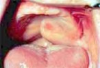
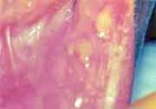
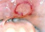
Adenoid Cystic Carcinoma of the left hard and soft palate. The lesion is poorly circumscribed. It is fixed to both superficial and deep structures.
|
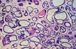 |
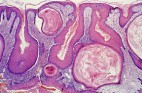
In this view, we can appreciate the ductal spaces with their pink product contrasting with paler material in the others
|
 |
This is the typical cribriform pattern of tumor cells in adenoid cystic carcinoma. There are small spaces lined by ductal cells and larger spaces containing basement membrane-like material. The pattern overall resembles a slice of Swiss cheese.
|
|

|
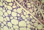
At Hi Mag, the duct lining cells are visible in small clusters with pink cytoplasm. The stellate cells surrounding the ducts and the larger round spaces are myoepithelial variants.
|
|
Congenital keratotic cyst
|
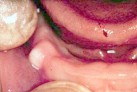 |
Congenital keratotic cyst, also known as dental lamina cyst. This lesion is a white, smooth surface lesion which is present on the alveolar ridge of neonates. It resolves spontaneously.
|
 |
In this image of gingiva from a newborn, we see numerous tiny keratotic cysts, some of which are merging with surface epithelium. Presumably, most or all such dental lamina remnants disappear this way without treatment
|
|
Aphthous Ulcers
|
|
|
Aphthous ulcers on the lower left labial mucosa. The ulcers are covered by a tan-yellow fibrin clot
|
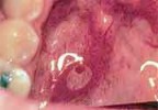 |
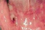
Aphthous ulcer on the left floor of the mouth.
|
 |
Aphthous ulcer on the right soft palate. Note the tan fibrin clot on the surface of the ulcer and the surrounding zone of erythema.
|
|
|
Large aphthous ulcer of the upper labial mucosa. This ulcer is much larger and of longer duration than typical aphthae
|
 |
Note the white areas of scarring on the labial mucosa. Some clinicians would call this major aphthae.
|
 |
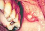
Aphthous ulcer of right lower labial mucosa, Ulcer is covered by a tan fibrin clot.
|
 |
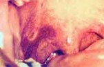
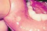
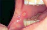
Aphthous ulcer on parotid papilla, where Stensen's duct enters the oral cavity.
|
 |
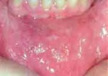
Several aphthous ulcers on lower right labial mucosa; there are also white areas of scarring closer to the midline. This patient has major aphthae.
|
 |
Photomicrograph of aphthous ulcer, the surface is covered by stratified squamous epithelium, with an ulcer in the center. There are no diagnostic microscopic findings of apthous ulcers. The diagnosis is based on the clinical findings and history
|
|
|
Aphthous ulcer; note the ulcer on the nonkeratinized epithelium of the buccal mucosa
|
 |
Aphthous ulcer on the nonkeratinized epithelium of the buccal mucosa
|


